What is it
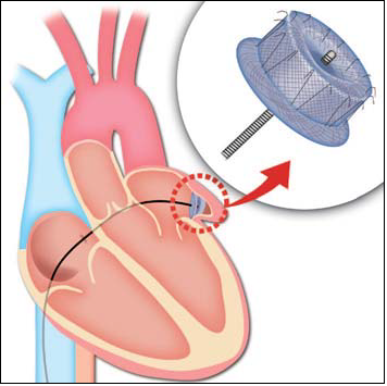
The left atrial appendage is a small cul-de-sac in the wall of the left atrium (the chamber that receives oxygenated blood from the lung). This is the most frequent localization of blood clot in the heart. Blood clots can occur when fast heart beats (atrial fibrillation or flutter) hinder proper contraction of the left atrial appendage. Blood therefore stagnates and coagulates, because it is no longer squeezed out from the appendage. Appendage occlusion avoids blood entering and clot formation. An “umbrella”- like device is implanted at the origin of the left atrial appendage, and seals its entrance, making it impossible for a clot to be created in the heart structure (Fig 5). A different option is to close the appendage from the outer side of the heart with a specific wire tightening the neck of the appendage. This is done once the outer heart, the so called “epicardial space” (comprised between the 2 epicardial sheets, and a thin layer of fibrous tissue that fixes the heart to the thorax and contains a small amount of fluid so that the heart can freely move) is reached with a puncture through the superior part of the abdominal wall. Patients are usually discharged 2 days after the operation.