ABLATION001 CS5


 . Catheter Ablation

What is it

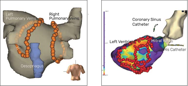
Catheter ablation is the technique during which catheters (small flexible tube) with electrodes (able to record and display the electrical activity of the heart on a monitor) are threaded up to the heart, in order to identify the area causing the tachycardia (fast heart beats). An ablation (a permanent damage) of the abnormally active heart cells is achieved with the delivery of heat or cold energy. Catheter ablation is performed in the chambers collecting (the atria) and pumping (the ventricles) blood with risk and success rates that vary depending from the specific arrhythmia. Nowadays, so called three-dimensional mapping system are routinely used in order to improve efficacy and safety, allowing precise movements in the heart without the need to use fluoroscopy, therefore dramatically reducing X-ray exposure (Fig. 6&7). Patients are usually discharged 2 days after the operation.


What we perform

All currently available 3D-Mapping Systems are used to treat the full spectrum of arrhythmias:

Ablation of simple (AV-Node-Reentrant-Tachycardia, AV-Reentrant-Tachycardia, Atrial Tachycardia, Typical Atrial Flutter) and complex (Atypical Atrial Flutter and Atrial fibrillation, Fig. 6) atrial arrhythmias

Ablation of ventricular arrhythmias in structurally normal and abnormal hearts (Fig. 7) with epicardial access if needed Ablation in congenital heart disease.

Immagine catetere

Why a patient needs a catheter ablation

Tachycardia originates from atria or ventricles. Palpitations, anxiety, shortness of breath, and an inability to exercise are usual symptoms. Syncope can occur, and tachycardias originating from the ventricle (ventricular tachycardia or fibrillation) can lead to cardiac arrest. In addition, a persistent high heartbeat can damage the heart resulting in a specific form of heart failure (tachycardiomyopathy). Treatment of the arrhythmia leads to symptom resolution or quick normalization of the heart function.

Catheter Ablation